
Development of a system for the analysis and classification of blood and bone marrow cell images to support morphological diagnosis of leukemia
Abstract
The University of Kaiserslautern and the Westpfalz-Klinikum have undertaken an interdisciplinary project for the microscopic analysis of blood and bone marrow cells using digital image processing for the early diangosis of leukemia.
Description of Project
After the microscopic blood and bone marrow images are acquired, the
cells have to be segmented, that is seperated from the background. They
also have to be divided into cytoplasm and nucleus. If some cells are clustered
together, they have to be seperated using a suitable algorithm since an
automatic analysis of the cells is not possible otherwise. Cellclusters
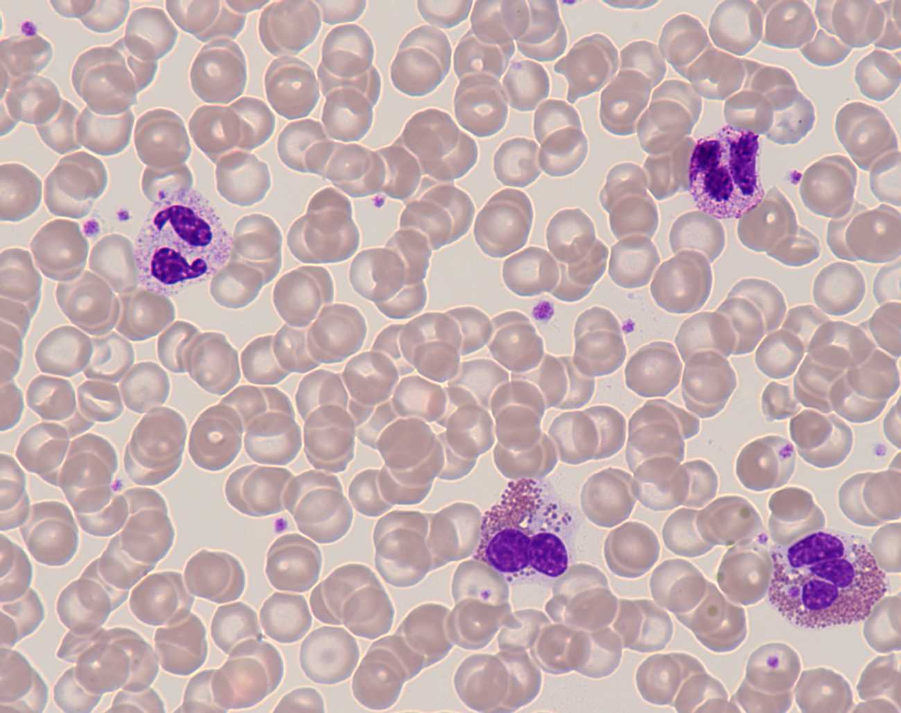
are usually found in bone marrow smears. In blood smears the cells are
usually already seperate or appear in loose groups of only a few cells.
The image on the left side shows a typical blood smear, the image on the
right a typical bone marrow smear.
|
Blood Smear |
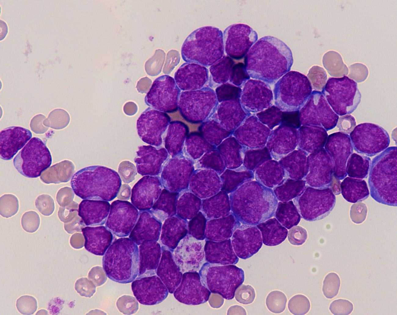
Bone Marrow Smear |
Once the cells are seperate, feature extraction can be taken up. The features are algorithmic realisations of verbal descriptions by physicians - for example the verbal description "finely woven" can be quantified via texture analysis. These quantified features are part of a feature vector which can then be used as a basis for classification. Among others, features from the following feature-spaces have been examined: geometric features, form oriented features, texture features, color features and combined features.
The essential steps of the overall project are:
- Setting up a data bank for the acquisition of microscopic images of blood cells
- Standardising the description of the blood cell images using formalised languages
- Development of an automatic image acquisition process
- Standardising the image formats and the process of archiving and retrieval of images
- Image analysis of the blood cell images for finding quantitative descriptive features of the bood cells is the core issue. This includes analysis, quantification, extraction and selection of features of the cell images. The correlation (i.e. the reationship) of the parameters of the features with the diagnosis of the physician has to be established. Once this relationship has been determined, it can be used as a tool for future diagnosis. The image analysis consists of the following:
- Color transformation from RYB to HSI (Hue, Saturation, Intensity)
- Segmentation methods to separate cells from background
- Declustering of clusters of blood cells
- Feature analysis of geometric shapes of the blood cells to obtain feature vectors
- Geomtric features: Cell dimensions, invariant moments of 1st ot 4th order, area ratio of cytoplasm and nucleus, number of segments in the nucleus
- Form oriented features: Cuvedness of the contours, contour energy of nucleus and cytoplasm
- Texture features: Entropy, contrast, homogenity, correlation, roughness, orientation, degree of orientation, busyness, Gabor features, Watershed features
- Color features: Hue and saturation of the cytoplasm
- Combined color and texture features (currently being implemented): Granularity in cytoplasm and their colors, stick-like form (in nucleus)
- Correlation of feature vectors with diagnosis of mutilated and immature blood cells
- Histogram analysis of mutilated and immature blood cells
Future Work and Work in Progress
- Classification via Neural Network
- Conceptual Graphs
- Feature Extraction
- Genetic Algorithms for Classification
- and others
Cooperations
Publications
M. Pandit, H. Hengen, Image Analysis of Blood and Bone Marrow Smears, IEEE/BMESI, BIOVISION, Bangalore, 2001
M. Pandit, H. Hengen, H. Link, F.-G. Hagmann, Computergestützte Diagnose von Leukämien unter Anwendung von Verfahren der digitalen Bildverarbeitung, DGHO Mannheim, 2001
T. Heger, H. Hengen, M. Pandit, Bildverarbeitung für Klassifikationsaufgaben in der Medizin und Qualitätssicherung, Automatisierungstechnik (at), Oldenbourg Verlag, 2002
H. Hengen, S. Spoor, M. Pandit, Analysis of Blood and Bone Marrow Smears using Digital Image Processing Techniques, SPIE Medical Imaging, San Diego, Feb. 2002
Finished Studienarbeiten, Diplomarbeiten, Project-Thesis, Master-Thesis
Objektorientierte Systemanalyse und -synthese in der digitalen Bildverarbeitung
(Object oriented systemanalysis and -synthesis for digital image processing)
Studienarbeit, Universität Kaiserslautern, 1997
O. Gabel:
Segmentation of Blood Cells
Projektarbeit, Indian Institute of Science, Bangalore, 1999
A. Hajra:
Segmentation and Feature Analysis of Blood Cells
Master Thesis, Universität Kaiserslautern, 2000
T. Paulus:
Entwurf und Realisierung einer Plattform zur Akquisition, Analyse
und Segmentierung von mikroskopischen Aufnahmen für die midizinische
Bildverarbeitung
(Design and implementation of a platform for acquisition, analysis
and segmentation of microscopic images)
Diplomarbeit, Universität Kaiserslautern, 2000
M. Ross:
Methoden der digitalen Bildverarbeitung für die Leukämiediagnostik
(Methods of digital image processing for Leukemia diagnosis)
Diplomarbeit, Universität Kaiserslautern, 2001
S. Spoor:
Normalisierung, Declustering und Merkmalsextraktion in der medizinischen
Bildverarbeitung
(Implementation of Image Standardisation and Advanced Segmentation
and Feature Extraction Procedures)
Diplomarbeit, Universität Kaiserslautern, 2002
Download
| Contact: | Susanne L. Siegrist | ||
| Room: | 12/324 | ||
| Phone: | 0631 / 205-2820 | ||
| e-mail: | |||
| or: | Heiko Hengen | ||
| Room: | 12/347 | ||
| Phone: | 0631 / 205-2091 | ||
| e-mail: |